
Deuteranomaly (Green-Weak): The Most Common Color Blindness
When someone says they are "color blind," statistically they most likely have deuteranomaly. It affects roughly 5% of men — more than every other type combined. The key point: the green cone isn't missing, its peak sensitivity is simply shifted.
~5%
Prevalence (Men) — highest
M
Shifted Cone (Green, not missing)
3
Working cone types (trichromat)
Deuteranomaly is a red-green color vision deficiency in which the green-sensitive M-cones are still present, but their spectral peak is shifted toward the L (red-sensitive) cones. It is the most common color vision deficiency of all, affecting roughly 5% of men. Because all three cone types are still working, a person with deuteranomaly is technically still a trichromat — just an anomalous one.
That distinction matters enormously, because it is what separates deuteranomaly from deuteranopia (green-blind). Deuteranopia removes an entire dimension of color vision; deuteranomaly merely compresses it. How much it is compressed varies enormously from person to person.
Colors commonly confused
Green ↔ yellow/orange, pink ↔ gray, cyan ↔ gray, plus many desaturated red-green pairs. How badly these confuse depends on severity — a mild deuteranomal may only slip up when colors are pale, small, or poorly lit.
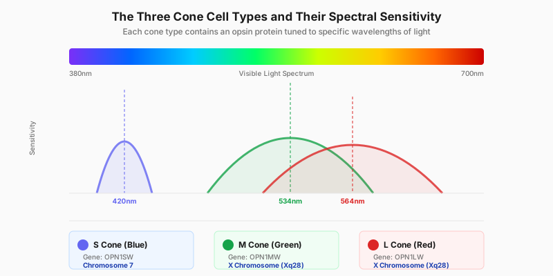
In a normal eye the M-cone peaks near 534 nm and the L-cone near 564 nm. Deuteranomaly pushes the M peak toward the L peak, the two curves overlap more, and the distinguishable difference between red and green shrinks.
How Deuteranomaly Works: Shifted, Not Missing
Human color vision runs on three cone types: L (long-wavelength / red, peaking near 564 nm), M (medium-wavelength / green, peaking near 534 nm), and S (short-wavelength / blue, near 420 nm). The brain works out color by comparing the relative strengths of those three signals — and in particular, the small difference between L and M carries the entire red-green axis.
In deuteranomaly, the M-cone photopigment is altered so that its peak sensitivity moves toward longer (redder) wavelengths. The closer the M peak sits to the L peak, the more the two response curves overlap, and the smaller the L-minus-M difference becomes. A smaller difference means a weaker signal for telling red from green — not an absent one. That is exactly why the condition is named with "-anomaly" (weak) rather than "-opia" (blind).
The gene involved is OPN1MW, which encodes the M-cone photopigment. In deuteranomaly it has typically recombined with the neighbouring OPN1LW gene, producing a hybrid pigment whose absorption peak lands somewhere between a normal M and a normal L.
"-anomaly" vs "-opia": One Suffix, Two Different Conditions
Color vision deficiencies are named by a strict rule, and reading the suffix tells you what you are dealing with. The prefix names which cone is involved (prot- = L/red, deuter- = M/green, trit- = S/blue). The suffix names what happened to it:
- -anomaly (weak) — the cone is present but its photopigment peak is shifted. Three usable cone types remain, so the person is an anomalous trichromat and keeps partial red-green discrimination. Deuteranomaly sits here.
- -opia (blind) — the cone is entirely absent. Only two usable cone types remain, making the person a dichromat, with that color axis essentially gone. Deuteranopia sits here.
So deuteranomaly and deuteranopia are not two names for one condition — they differ at the level of the receptor itself: one weakens a signal, the other deletes it. It also explains why someone with deuteranomaly gets colors right "sometimes" on charts, indicator lights, and fruit, while a deuteranope gets them wrong consistently.
Deuteranomaly Is a Spectrum, Not a Switch
This is the most misunderstood thing about deuteranomaly. Dichromacy, like deuteranopia, is essentially binary — the cone is there or it isn't. Deuteranomaly's severity instead depends on how far the M-cone's peak has actually shifted, and that shift varies continuously. Two people with the same diagnosis can have very different day-to-day experiences:
- Mild — a small shift. Almost nothing is noticeable day to day; mistakes creep in only with pale colors, tiny color patches, or dim light. Many of these people never find out they have it until a medical screening or an occupational color vision test.
- Moderate — a clear shift. Green versus yellow-orange and pink versus gray get confused regularly, clothing choices and red-green charts become a recurring nuisance, but large, saturated reds and greens are still told apart.
- Severe — the shift brings the M peak close to the L peak, and the two responses nearly coincide. The lived experience here approaches deuteranopia, with the red-green axis largely collapsed, even though the person is still physiologically an anomalous trichromat.
This spectrum is why "am I actually color blind?" is such a hard question to answer for deuteranomaly — and why an Ishihara test cannot grade it and an anomaloscope can, which we cover below.
How It Affects Everyday Vision
The world does not look gray or straightforwardly "wrong" to someone with deuteranomaly. Blues and yellows stay vivid, and reds keep their normal brightness — unlike protanopia, where reds are additionally darkened. What gets compressed is the resolution along the red-green axis.
Typically greens drift toward yellow or orange, pinks are hard to separate from grays, and cyan can read as gray. More subtly, the paler, smaller, and dimmer the color, the more likely the error — two large saturated blocks of red and green side by side may be no trouble at all, while the same two colors shrunk into a chart legend become indistinguishable. That inconsistency is why people with deuteranomaly often doubt their own experience.
Causes and Genetics
Deuteranomaly is almost always inherited. The OPN1MW gene sits on the X chromosome (locus Xq28), arranged in an array right next to OPN1LW — and it is precisely that neighbouring arrangement that makes unequal crossing over and recombination likely, producing hybrid pigments whose peak falls between the two. That is the molecular basis of deuteranomaly.
Inheritance is X-linked recessive. Males have a single X chromosome, so one altered copy is enough to show the trait; females have two X chromosomes and need both altered, with a single copy making them carriers. That is why deuteranomaly is far more common in men than in women. If the mother is a carrier, each son has a 50% chance of being deuteranomalous and each daughter a 50% chance of being a carrier. For a deeper look, see our genetics of color blindness guide.
How Deuteranomaly Is Diagnosed
Diagnosing deuteranomaly means answering two separate questions: is it there? (screening) and how severe is it? (grading). Different tools are good at different questions:
- Ishihara test — the standard screen. It will pick deuteranomaly up, but it cannot grade severity: it tells you a red-green deficiency exists, not how far the cone has shifted. Very mild deuteranomaly can pass the whole plate set.
- Red-green color blind test — a quick targeted self-check of the red-green axis, good as a first step.
- Farnsworth D-15 test — an arrangement test that shows the confusion axis, separates deutan from protan, and broadly splits mild from moderate-severe.
- FM100 Hue test — the most granular arrangement test, scoring overall hue discrimination ability.
- Anomaloscope — the gold standard for grading. The subject adjusts a mixture of red and green light to match a fixed yellow. Someone with deuteranomaly accepts a green-shifted mixture ratio, and accepts a wider range of ratios (a wider matching range) than a normal observer. The direction of the shift confirms it is deutan; the width of the matching range gives the severity — a quantitative result no other test provides.
Daily Life Impact (5 Concrete Scenarios)
- Charts and dashboards — red-green line charts, transit maps, and red/green conditional formatting in spreadsheets are the most-cited frustration, because legend swatches are small and desaturated.
- Choosing and matching clothes — pink versus gray, cyan versus gray, and green versus brown go wrong easily, especially under a shop's warm lighting.
- Judging ripeness — the transitional shades between a green and a yellow banana, or an unripe and a half-ripe tomato, are the hardest calls, because the difference lies exactly on the compressed red-green axis.
- Status indicator LEDs — the tiny red/green LEDs on chargers, routers, and power supplies are the classic failure case; blink pattern or position is a more reliable cue than color.
- Occupational color standards — most jobs are unaffected, but electrical wire identification, aviation, parts of the military, and some rail and marine roles have explicit color vision requirements, usually enforced with a named test. See our guide to jobs for colorblind people.
Tips and Adaptation
- Ask for charts to encode data redundantly with shape, line style, and direct labels, not red versus green alone.
- A phone color-picker app names an object's true color instantly — genuinely useful when buying clothes or sorting wires.
- Built-in OS colorblind filters and high-contrast modes noticeably improve small on-screen swatches.
- Choose colorblind-safe palettes (Wong, Viridis) over red-green contrasts, and make them your team's default.
- Judge colors in good light and on large patches — the same color is far easier to call correctly when the area is bigger.
- Corrective glasses (e.g. EnChroma) help some anomalous trichromats subjectively, but results vary and they are not a cure — see how color blind glasses work.
When to See a Doctor
Inherited deuteranomaly is stable, lifelong, needs no treatment, and does not get worse. What does warrant prompt attention is the opposite pattern: color vision that changes suddenly, affects only one eye, or comes alongside reduced acuity or visual field loss. That points to a retinal or optic-nerve problem rather than an inherited deficiency.
Two other situations are worth a formal assessment. First, a child who keeps getting colors wrong while learning them — an early diagnosis lets teachers adapt classroom materials; see early signs of color blindness in children. Second, if you are heading into a field with color vision standards, getting a formal result early beats discovering it at the assessment.
Frequently Asked Questions
Sources
- National Eye Institute (NEI) — Overview of color blindness types and prevalence
- American Academy of Ophthalmology (AAO) — Clinical information on red-green color vision deficiency
- Colour Blind Awareness — Anomalous trichromacy and deuteranomaly confused-color list
- National Health Service (NHS) — Causes, diagnosis and management of colour vision deficiency
- MedlinePlus Genetics — OPN1MW/OPN1LW genes and X-linked inheritance
Take the Red-Green Test
Screen your red-green vision in 2 minutes
View Simulation
See the world through deuteranomaly
Related Types
- Deuteranopia (Green-Blind) — the same deutan family, but the M-cone is entirely absent (dichromacy)
- Protanopia (Red-Blind) — red-green blindness with missing L-cones; reds are additionally darkened
- Tritanopia (Blue-Blind) — rare blue-yellow deficiency, often acquired
- All 8 types of color blindness — overview