
Tritanomaly (Blue-Weak): A Weakened Blue-Yellow Axis, Not a Missing One
Tritanomaly is a mild blue-yellow deficiency in which the blue-sensitive S-cones are still there but spectrally shifted. Two things set it apart from red-green types: it is not X-linked, so men and women are affected equally — and it is more often acquired than inherited.
<1%
All tritan deficiencies
Equal
Sex ratio (not X-linked)
S
Shifted cone (blue, not missing)
Tritanomaly is a blue-yellow color vision deficiency in which the blue-sensitive S-cones are still present but their peak spectral sensitivity is shifted. Because all three cone types are still working, a person with tritanomaly remains a trichromat — just an anomalous one: signals along the blue-yellow axis arrive compressed and distorted rather than missing entirely. That single fact accounts for every experiential difference between tritanomaly and tritanopia (blue-blind).
Deficiencies on the blue-yellow axis — clinically called tritan defects — together affect well under 1% of people, far fewer than red-green types. That rarity is also why tritanomaly is so often missed, or written off as tired eyes or a badly calibrated screen.
Colors commonly confused (blue-yellow, mild)
Blue ↔ green, yellow ↔ pink, yellow ↔ light gray, violet ↔ dark red. Unlike tritanopia, someone with tritanomaly usually names strong, saturated blues and yellows correctly; errors cluster in pale, desaturated, small, or dimly lit samples. Red-green discrimination is entirely normal — which is why standard Ishihara plates show no errors at all.
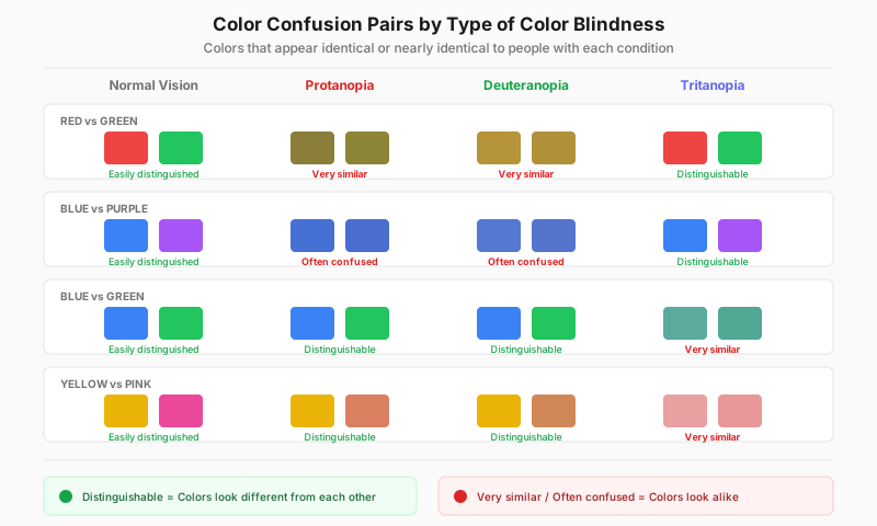
Tritanomaly affects the blue-yellow pairs in the lower rows — but more mildly than tritanopia: the pairs blur together rather than merging outright.
What Tritanomaly Actually Is
Human color vision relies on three cone types: L (long-wavelength / red), M (medium / green), and S (short / blue). A normal S-cone peaks in sensitivity around 420 nm, in the short-wavelength blue region. In tritanomaly the S-cone photopigment is altered by a genetic variant so that its sensitivity peak is shifted away from where it should be, and its response curve no longer lines up with the normal one.
The brain decides "blue or yellow?" by subtracting the S-cone signal from the combined L and M signal — the blue-yellow opponent channel. When the S response curve shifts, the result of that subtraction is compressed: two colors that should sit far apart on the axis now produce a much smaller difference, so subjectively they "look about the same." No dimension of color is lost — the resolution along one dimension is reduced.
One anatomical factor makes this worse. S-cones are by far the least numerous cone type in the retina, and the very center of the fovea contains almost none. Starting from such a small population, any weakening is easier to notice — especially in dim light and on small targets.
Tritanomaly vs. Tritanopia: One Suffix, Two Conditions
The two names get used interchangeably, but clinically they are different things. They share the same color axis; what differs is the state of the S-cone:
- Tritanomaly (blue-weak) — the S-cones are present with a shifted peak. All three color dimensions remain, so this is anomalous trichromacy. Blue-yellow discrimination is reduced, mildly to severely.
- Tritanopia (blue-blind) — the S-cones are absent or nonfunctional. Only two dimensions remain, so this is dichromacy, and the blue-yellow axis is effectively gone.
A practical self-check: if you have no trouble at all with strong, saturated blue versus yellow, and only go wrong on near-neighbors like pale blue versus gray or pale yellow versus pink, that points toward the "weak" form rather than the "blind" one. Real grading requires an arrangement test — see the diagnosis section below. For the severe form in full, read our tritanopia guide.
How It Affects Everyday Vision
Nothing is missing from a tritanomalous world — reds, oranges, and greens all look normal, and red-green discrimination is completely intact. The changes sit at the short-wavelength end of the spectrum: the blue of sky and water can read as greenish or gray; pale yellow slides toward pink or light gray under some lighting; the line between violet and dark red blurs; and the whole cyan-teal-turquoise family collapses into one band.
Because these pairs rarely carry safety-critical information — traffic lights and warning palettes all live on the red-green axis — tritanomaly tends to be harmless to safety but persistently disruptive to judgment. Matching clothes, picking a paint chip, or reading a blue-yellow chart means guessing wrong repeatedly with nobody there to correct you. Plenty of people only find out in adulthood, after happening to take a blue-yellow test.
Causes and Genetics: Why Men and Women Are Affected Equally
This is the biggest structural divide between tritanomaly and the red-green types. The red-green opsin genes (OPN1LW and OPN1MW) sit on the X chromosome and are inherited in an X-linked recessive pattern. Men have a single X, so one defective copy is enough — which is why red-green color blindness is overwhelmingly male.
Tritanomaly does not work that way. The S-cone opsin gene OPN1SW sits on chromosome 7 — an autosome, not a sex chromosome. Inherited tritan deficiency is passed down in an autosomal dominant pattern with incomplete penetrance. Three consequences follow directly:
- Men and women are affected at equal rates — there is no male skew here, and that absence is itself a useful clue that a deficiency belongs to the tritan family rather than the red-green one.
- Either parent can pass it directly to a child of either sex — no carrier-mother pathway of the kind X-linked traits require.
- Severity varies widely within one family — incomplete penetrance means relatives carrying the same variant can range from nearly asymptomatic to close to tritanopia.
For a systematic comparison of inheritance patterns across all types, see our genetics of color blindness guide.
Acquired Is More Common Than Inherited — the Key Clinical Fact About Tritan Defects
Red-green color blindness is almost always congenital and stable for life. The blue-yellow axis is different: acquired blue-yellow deficiency is more common than the inherited kind. The blue-yellow channel is simply fragile — few S-cones, and a pathway that is unusually sensitive to retinal and optic-nerve damage — so it is frequently the first axis to degrade. Common acquired causes include:
- Glaucoma — damages the optic nerve; loss of blue-yellow discrimination is regarded as one of the earlier functional changes.
- Diabetic retinopathy — reduced blue-yellow discrimination can appear before any obvious change in acuity.
- Optic neuropathies — inflammatory, ischemic, and toxic forms among them.
- Age-related yellowing of the lens — the lens grows progressively yellower with age and absorbs more short-wavelength blue light, producing a gradual tritan-like shift. This is normal aging, but it stacks on top of any existing deficiency.
- Age-related macular degeneration and other retinal disease.
- Certain medications and industrial solvents — some drugs and organic-solvent exposures affect color vision, and the blue-yellow axis is often hit first.
The takeaway is blunt: a blue-yellow color problem that appears suddenly is a warning sign, not something to dismiss as "always been that way." Congenital tritanomaly is present from childhood, symmetric in both eyes, and stable for life. Acquired deficiency usually has a datable onset, may affect one eye only, and tends to worsen over time.
How Tritanomaly Is Diagnosed
The most important point first: standard Ishihara plates cannot detect a tritan deficiency. They are built for the red-green axis, so a person with pure tritanomaly typically finishes with a perfect score — which is exactly why blue-yellow deficiency goes undiagnosed for so long. Detecting it requires tools aimed at the blue-yellow axis:
- Blue-yellow color blind test — plate screening designed for the tritan axis; the first test to take if you suspect tritanomaly.
- Farnsworth D-15 test — an arrangement test. You order color caps by hue, and the direction of the crossing lines on the result plot shows directly whether the confusion axis is tritan or red-green.
- FM100 Hue test — the most detailed arrangement test; it identifies the axis and quantifies severity, which is what separates "weak" from "blind" and makes it useful for retesting over time to monitor an acquired change.
- Anomaloscope with a Moreland match — the clinical gold standard for confirmation, done at an eye clinic.
- A full eye exam — if an acquired cause is suspected, intraocular pressure, retina, and optic nerve all need checking.
If you are not sure which axis you are on, take an Ishihara test first to rule out a red-green problem, then a blue-yellow test. The combination of the two results is itself diagnostically informative.
Daily Life Impact (5 Concrete Scenarios)
- Clothes and color matching — navy versus charcoal, teal versus blue-green look nearly identical under store lighting; a pale yellow shirt can read as pale pink.
- Charts and maps — business dashboards lean heavily on blue-to-yellow gradients, and many "colorblind-safe" palettes deliberately swap blue-yellow in for red-green, which makes them the hardest charts of all for a tritanomalous reader.
- Screens and color work — white balance drifts during photo editing, so work looks too blue or too yellow on other people's displays; blue-green hierarchies in design are hard to control.
- Outdoors and nature — sky and sea blues can look greenish or washed out, and low-saturation colors at dusk are the hardest of all to judge.
- Health self-monitoring — for anyone with known diabetes, glaucoma, or optic-nerve disease, change in blue-yellow discrimination is a functional indicator worth rechecking periodically with the same test.
Tips and Adaptation
- Never encode information in blue versus yellow alone — add labels, patterns, shapes, or a brightness difference as a second channel.
- A phone color-picker app names an object's true color on the spot; most useful when buying clothes or choosing paint.
- Push saturation and brightness apart: the same two colors become far easier to separate once their saturation differs noticeably.
- OS-level colorblind filters and high-contrast modes help on screen; for color work, make the final call from numbers (HEX/HSL), not by eye.
- Prefer colorblind-safe palettes (Wong, Viridis) — but check them yourself, since many were designed for red-green types and lean on exactly the blue-yellow contrasts that fail here.
- If the deficiency is acquired, the priority is managing the underlying cause with your doctor (blood sugar, eye pressure, optic-nerve follow-up) rather than correcting color. On the limits of tinted lenses, see how color blind glasses work — they are designed mainly for red-green types and do little for tritan deficiencies.
When to See a Doctor
Because a larger share of blue-yellow deficiency is acquired, this section matters more for tritanomaly than for any red-green type. If any of the following apply, get examined rather than assuming you were simply born this way:
- Blue-yellow trouble that appeared suddenly, or that you can date to roughly a particular time.
- A difference between your two eyes — cover one eye at a time and look at the same object; congenital deficiency is symmetric, so a monocular difference strongly suggests acquired disease.
- Progressive worsening — the same test scored months apart keeps getting worse.
- Accompanying loss of acuity, visual field defects, eye pain, flashes, or new floaters.
- You have diabetes, a family history of glaucoma, or take a medication known to affect vision.
Catching an underlying cause early matters: damage from glaucoma and diabetic retinopathy is generally irreversible, but early treatment can stop further vision loss. Congenital tritanomaly, by contrast, needs no treatment at all — it is stable, non-progressive, and never leads to blindness.
Frequently Asked Questions
Sources
- National Eye Institute (NEI) — Overview of color vision deficiency types, including blue-yellow (tritan)
- American Academy of Ophthalmology (AAO) — Clinical information on blue-yellow deficiency and anomalous trichromacy
- MedlinePlus Genetics — OPN1SW — The S-cone opsin gene on chromosome 7 and its autosomal dominant inheritance
- Colour Blind Awareness — Distinguishing tritanomaly from tritanopia, and confused-color lists
- National Health Service (NHS) — Causes, acquired factors, and management of colour vision deficiency
Take the Blue-Yellow Test
Screen the blue-yellow axis (Ishihara can't)
Take the D-15 Arrangement Test
See whether your confusion axis is tritan
Related Types
- Tritanopia (Blue-Blind) — the severe form on the same axis: S-cones absent, a dichromacy
- Protanopia (Red-Blind) — red-green blindness with missing L-cones; X-linked and mostly male
- Deuteranopia (Green-Blind) — red-green blindness with missing M-cones; reds keep normal brightness
- All 8 types of color blindness — overview