
Protanomaly (Red-Weak): Red Is Still There, Just Dimmer
Protanomaly is not a missing cone. The red-sensitive L-cone is present but its peak sensitivity has drifted toward green. Red-green discrimination gets harder — and, more importantly, red itself gets dimmer, with real safety consequences.
~1%
Prevalence (Men)
3
Cone Types (still three)
L
Shifted Cone (Red)
Protanomaly is the milder branch of red-green color vision deficiency. Unlike protanopia (red-blind), the L-cones are not missing in protanomaly — they are present and functioning, but their peak spectral sensitivity is shifted toward that of the M (green-sensitive) cone. All three cone types are still there, so a person with protanomaly is still a trichromat — just an anomalous one.
That distinction shapes everything else on this page. Someone with protanomaly keeps partial red-green discrimination; their color world has not collapsed from three dimensions to two the way a protanope's has. Protanomaly affects roughly 1% of men and is less common than deuteranomaly, the green-weak equivalent.
Colors commonly confused
Red ↔ brown / dark green, orange ↔ yellow-green, purple ↔ blue, and desaturated pinks ↔ gray. Because red is also darkened, deep reds often read as an almost-black brown.
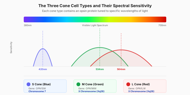
The three normal cone sensitivity curves. In protanomaly the L curve slides toward the M curve — the curve is still there, it is just standing in the wrong place.
Anomalous Trichromacy: Why It Is "Weak," Not "Blind"
Color vision deficiencies follow a naming convention, and once you know it, protanomaly explains itself. The suffix -opia means a cone type is functionally absent, leaving only two working cone classes — that is dichromacy. The suffix -anomaly means all three cone types are present, but one of them carries an altered photopigment whose peak sensitivity has moved. That is anomalous trichromacy.
Protanomaly is the second case. The L-cone photopigment is replaced by a hybrid version whose absorption peak no longer sits at the long-wavelength red end but has drifted toward the M-cone peak. When the L and M curves overlap too much, the two signals the brain subtracts to compute "how red versus how green" become nearly identical — the difference is flattened, and red-green color resolution drops.
How far the peak has moved varies from person to person, which is why protanomaly ranges from mild to severe. A mildly affected person may go a lifetime without noticing; a severely affected one approaches protanopia. Think of protanomaly and protanopia as different points on one spectrum of severity, not as two unrelated conditions.
Reduced Red Luminance: The Defining Feature
If you remember one thing about protanomaly, make it this: red is not merely the wrong hue, it is dimmer.
The L-cone does not only decide "the hue red" — it is the main contributor to how bright long-wavelength light appears. Once its peak drifts away from the red end, red light produces a weaker response, and the brain registers red at a lower luminance. Bright red looks dull and brownish; deep red comes close to black.
This is the biggest difference between protanomaly and deuteranomaly. In deuteranomaly it is the M-cone that is shifted, and the long-wavelength luminance channel is left essentially intact — so a deuteranomalous observer confuses the same hues but has no marked brightness loss; red still looks bright enough to them. Two people can fail the same color plate, and only the protan one will feel that the whole red region has sunk into shadow.
Clinically the loss is measurable. In the anomaloscope's Rayleigh match, a protanomalous observer not only needs an unusually red-heavy mixture, they also turn the red primary up in brightness before it looks matched — the key cue that identifies a protan deficiency.
Why "Only Mild" Can Still Mean Higher Real-World Risk
It is tempting to assume "weak" is a harmless version of "blind." For protan-type deficiencies that inference fails. Protanomaly discriminates colors better than protanopia does, but it carries the same reduced red luminance — and an enormous share of the world's safety signals are coded in red: brake lights, red traffic lights, machine fault indicators, fire equipment, hazard markings.
Those signals rely on being red and bright to grab attention. When red is perceived as dimmer, what is lost is not just the color information but detection itself: a car's brake lights coming on in the distance have less contrast and pop out later, and the gap widens at night or in rain and fog. This is where protanomaly and deuteranomaly genuinely diverge in practical risk. For the full discussion, see our guide to driving with color blindness.
How It Affects Everyday Vision
A person with protanomaly does not see a gray world — blues and yellows stay vivid, and most colors can still be named. What is compressed is the resolution along the red-green axis: nearby shades stop separating, rather than all color disappearing.
In practice, a red strawberry among green leaves does not jump out the way it does for someone with normal vision; red brick, terracotta, and brown wood all read as one family; the boundary between pink and pale gray, or purple and blue, gets blurry. Layer the darkening of red on top, and a lot of information that was designed to be the most conspicuous thing on screen becomes the least conspicuous.
Causes and Genetics
Protanomaly is almost always inherited. The OPN1LW gene for the L-cone photopigment sits right next to the OPN1MW gene for the M-cone pigment on the X chromosome, and the two sequences are highly similar. That similarity is exactly what makes them prone to unequal crossing-over and recombination during meiosis, producing hybrid L/M genes. When a hybrid gene encodes a pigment whose peak falls somewhere between the normal L and M peaks, the result is protanomaly.
Inheritance is X-linked recessive. Males have a single X chromosome, so one altered copy is enough to cause the condition; females have two X chromosomes and usually need both copies altered, which is why women are far more often carriers than affected. If the mother is a carrier, each son has about a 50% chance of being affected. For a deeper look at inheritance patterns, see our genetics of color blindness guide.
How Protanomaly Is Diagnosed
Mild protanomaly is one of the most easily missed deficiencies — plenty of people pass part of a plate test and only find out at an occupational screening as an adult. The sensible path is screen first, then classify and grade:
- Ishihara test — the first-line screen; flags red-green deficiency, but a mild anomaly can partly pass, and plates alone do not separate protan from deutan.
- Red-green color blind test — a quick targeted self-check of the red-green axis, good as a first step.
- Farnsworth D-15 test — an arrangement test whose pattern of misplacements reveals the confusion axis, helping separate protan from deutan.
- FM100 Hue test — the most detailed arrangement test; reflects overall color discrimination ability.
- Anomaloscope — the gold standard for both diagnosis and grading. Its Rayleigh match (a red-green mixture set against a yellow reference) yields two facts at once: how much red is needed for a match, which fixes the type and degree, and whether the red primary has to be turned up in brightness, which exposes reduced red luminance and confirms a protan deficiency.
Online tests give a genuinely useful signal, but screen calibration and ambient light both affect the result — treat a clinical exam as the authority on formal classification.
Daily Life Impact (5 Concrete Scenarios)
- Driving and brake lights — distant brake lights and red signals have too little contrast and register later, worst at night and in rain or fog. Reading a signal by position (red on top) beats reading it by color.
- Judging cooked food — the pink center of a steak versus fully browned meat is hard to call; ripeness of strawberries, tomatoes, and peppers needs touch and smell as backup.
- Device status LEDs — red/green bicolor indicators (charging vs. charged, OK vs. fault) are near-impossible to tell apart by color alone, and the red state looks dimmer than it really is.
- Charts and dashboards — red-green gauges, profit/loss tables, and heatmaps put red in a state that is both wrong-hued and darkened, so it is easily misread as a dark neutral.
- Clothing and color matching — red vs. brown, pink vs. gray, and purple vs. blue pairings go wrong; dark red garments frequently read as black under indoor lighting.
Tips and Adaptation
- Read signals by position and order, never by color — traffic lights, indicator LEDs, progress bars.
- A phone color-picker app names an object's true color on the spot; most useful when buying clothes or furnishings.
- Build reduced red luminance into driving habits: leave a larger following distance and do not treat "I saw the brake lights" as your only cue.
- OS-level colorblind filters and high-contrast modes improve red-green separation on screen.
- When making charts, switch to colorblind-safe palettes (Wong, Viridis) and encode redundantly with shape or labels instead of relying on red-green.
- Corrective glasses (e.g. EnChroma) deepen the separation between the L and M channels and help some anomalous trichromats subjectively, but results vary and they cannot restore the lost brightness information — see how color blind glasses work.
When to See a Doctor
Inherited protanomaly is stable, lifelong, and needs no treatment in itself. Three patterns do warrant a visit: color vision that changes suddenly or keeps worsening, a deficiency affecting only one eye, and color trouble accompanied by reduced acuity, visual field loss, or light sensitivity. Those point to acquired color vision problems of the retina, optic nerve, or medication — not inherited color weakness — and should be assessed by an eye-care professional promptly.
Separately, if you are heading toward a career with hard color vision requirements (aviation, some military and police roles, rail, electrical work), get a formal classification done early rather than discovering the result at a pre-employment exam.
Frequently Asked Questions
Sources
- National Eye Institute (NEI) — Overview of color blindness types and prevalence
- American Academy of Ophthalmology (AAO) — Clinical information on red-green color vision deficiency
- Colour Blind Awareness — Anomalous trichromacy confused-color list and inheritance
- National Health Service (NHS) — Causes and management of colour vision deficiency
- MedlinePlus Genetics — OPN1LW/OPN1MW genes and X-linked inheritance
Take the Red-Green Test
Screen your red-green vision in 2 minutes
View Simulation
See the world through protanomaly
Related Types
- Protanopia (Red-Blind) — the severe end of the same protan spectrum: L-cones functionally absent, a dichromacy with essentially no red-green discrimination
- Deuteranopia (Green-Blind) — red-green blindness with missing M-cones; reds keep normal brightness
- Tritanopia (Blue-Blind) — rare blue-yellow deficiency, often acquired
- All 8 types of color blindness — overview