
Achromatopsia (Total Color Blindness): A World of Light and Dark
Achromatopsia is not a confusion between two hues — it is a failure of the cone system as a whole. Vision runs on rods alone, so it brings not just grayscale sight but severe photophobia, nystagmus, and low visual acuity, setting it apart from every red-green or blue-yellow type.
~1 / 30,000
Global Prevalence
Equal M / F
Autosomal Recessive
L M S
All Cones Non-Functioning
Achromatopsia, also called total color blindness or rod monochromacy, is a condition in which all or nearly all of the retina's cone cells fail to function, leaving vision to run almost entirely on the rods. The result is a world seen only in shades of gray from black to white — no hue, no saturation, only brightness. It affects roughly 1 in 30,000 people worldwide, making it one of the rarest color vision disorders.
It is a fundamentally different thing from the "one cone type is missing" conditions such as protanopia and deuteranopia, and the section below on that difference is the most important part of this page.
What the world looks like
Every color collapses into grayscale: a red apple and a green apple differ only in lightness; a traffic light can only be read by which lamp is lit; a chart that codes data by hue alone conveys nothing. The image also runs bright and "overexposed," detail is not sharp, and outdoors in strong sun it becomes nearly impossible to see.
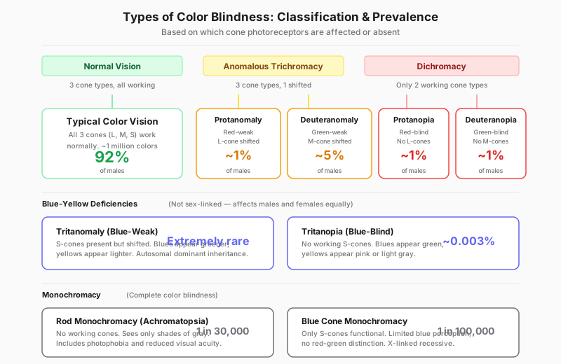
Achromatopsia sits at the far end of the spectrum: not a damaged color axis, but an absent color system.
What Achromatopsia Actually Is
The human retina holds two classes of photoreceptor. Cones handle color and fine detail in bright light and come in three types (L red, M green, S blue). Rods carry a single photopigment, are extremely sensitive but colorblind by nature, and handle vision in dim light.
In achromatopsia the cones' phototransduction pathway is broken — mutations in the genes controlling cone ion channels or transduction proteins prevent cones from converting light into a neural signal. The cones may still be structurally present, but functionally they are mute. Day and night alike, only the rods are reporting.
Because rods carry just one photopigment, the brain receives a single dimension of information: brightness. To distinguish two colors, the visual system needs at least two photoreceptor types with different response curves to compare against each other. With only one class left, color vision is physically impossible — which is where the name monochromacy comes from.
Why Achromatopsia Is Categorically Different: Photophobia, Nystagmus, Low Acuity
This is the key section of the page. Protanopia, deuteranopia, and tritanopia are hue-discrimination problems — visual acuity, light tolerance, and eye movement are all normal, and many people only discover their deficiency in adulthood when they finally take a test. Achromatopsia is nothing like that. Because what is lost is the entire daylight photoreceptor system, it necessarily brings three extra symptoms, often described together as the achromatopsia triad.
1. Photophobia and day blindness. Rods are built for dim light and saturate quickly under bright light. In a normal eye, cones take over as light levels rise; in achromatopsia the cones never take over, so in daylight the rods are flooded and the person is effectively dazzled — a state called hemeralopia, or day blindness. Many people with achromatopsia see best on overcast days, indoors, or at dusk.
2. Nystagmus. Cones are packed densely into the foveal center, and that is what the eye uses to lock onto a fixation target. With the cones not working, the fovea cannot supply stable fixation feedback, and the eyes drift back and forth involuntarily. This usually shows up in early infancy and is often the first clue that leads to diagnosis.
3. Reduced visual acuity. Fine detail depends on the dense cone mosaic of the fovea. Rods have far lower spatial resolution and are not concentrated at the foveal center, so best-corrected acuity in achromatopsia is typically well below normal and falls into the low-vision range. Glasses cannot fix it, because the problem is not refractive — it is in the photoreceptors themselves.
In one line: if someone "sees no color" but has normal acuity, no light sensitivity, and steady eyes, it is almost certainly not achromatopsia — it is a red-green or blue-yellow deficiency. These three companion symptoms are what clinicians use to tell them apart.
How It Affects Everyday Vision
People often imagine achromatopsia as "watching a black-and-white movie," but the lived experience is more complicated. First, the image is not a nicely balanced monochrome photograph — it is closer to an overexposed one, where large areas outdoors wash out to white and detail drowns in the light.
Second, anything that encodes information by hue alone fails completely: traffic lights, map legends, color-highlighted text, red/green status buttons, multiple colored lines on a chart. And the blue-yellow axis that a red-green dichromat can still fall back on is not available here either.
Third, low acuity stacked on top of photophobia makes reading, seeing a whiteboard, and recognizing faces much harder than a pure color problem would. Functionally, achromatopsia behaves more like a low-vision condition than a color vision one — which is why support and accommodations should be modeled on low-vision practice.
Causes and Genetics
Complete achromatopsia is autosomal recessive. It is caused by mutations in genes governing cone phototransduction — most commonly CNGB3, CNGA3, and GNAT2, along with PDE6C and PDE6H. These genes sit on ordinary autosomal chromosomes, not on the X chromosome.
That is the exact opposite of red-green color blindness, and the distinction matters. The OPN1LW and OPN1MW genes behind red-green deficiency are on the X chromosome and inherited X-linked recessively, which is why men are affected far more often than women. Achromatopsia is autosomal recessive: a child must inherit one mutated copy from each parent to be affected, and the parents are usually unaffected carriers. Because the sex chromosomes are not involved, men and women are affected at essentially equal rates. For a fuller treatment of inheritance patterns, see our genetics of color blindness guide.
One caveat: a rarer related condition called blue-cone monochromacy, in which only the S-cones retain function, produces similar but somewhat milder symptoms and is X-linked. It is the one X-linked exception among the monochromacies and should not be confused with complete achromatopsia.
Pingelap: Achromatopsia's Most Famous Case
The Pingelap atoll in Micronesia, in the Pacific, has one of the highest known concentrations of achromatopsia anywhere in the world — roughly one in ten islanders (~10%), against a global average of about 1 in 30,000.
The reason is a textbook founder effect. A devastating typhoon swept the island in the late 18th century and left only a handful of survivors, one of whom carried the recessive mutation. As the population rebuilt from that tiny gene pool — on a relatively isolated island with a high rate of marriage within the community — the recessive variant became common and homozygous (affected) individuals appeared in large numbers.
Neurologist Oliver Sacks documented the island in his book The Island of the Colorblind, which brought this rare condition to a general audience for the first time. Pingelap is also a standard genetics-textbook illustration of the founder effect.
How Achromatopsia Is Diagnosed
Diagnosis looks very different from red-green color blindness. It is usually not first spotted by a plate test — instead a baby is brought in for nystagmus and obvious light sensitivity, and the color loss is uncovered from there.
- Infant clinical clues — nystagmus appearing within the first months of life, unusual sensitivity to bright light, and poor visual following are the earliest signals.
- Electroretinography (ERG) — the core test. The classic achromatopsia pattern is a severely reduced or absent photopic (cone) response with a broadly normal scotopic (rod) response. That "cones gone, rods intact" signature is the key evidence.
- Genetic testing — finding biallelic mutations in CNGB3, CNGA3, GNAT2 and related genes confirms the diagnosis and matters for genetic counseling and for eligibility in gene therapy trials.
- Ishihara test — failed comprehensively, but it is a red-green screen and cannot separate total color blindness from severe red-green deficiency; it only signals that further workup is needed.
- FM100 Hue test and D-15 test — arrangements come out essentially random with no consistent confusion axis, which is itself suggestive of monochromacy.
- Acuity and fundus examination — to document best-corrected acuity and rule out other retinal disease.
An online plate test is fine as a first look, but suspected achromatopsia must be confirmed by an eye doctor with ERG and genetic testing. A web test cannot do it.
Daily Life Impact (5 Concrete Scenarios)
- Being outdoors on a sunny day — bright sun makes it nearly impossible to keep the eyes open and see; midday is worst. Many people wear dark filter lenses constantly or shift outings to early morning, dusk, or overcast days.
- Classroom and reading — low acuity plus photophobia makes whiteboards, projected slides, and small print hard; enlarged materials, electronic magnifiers, front-row seating, and dimmable lighting are typical needs.
- Driving — unlike red-green color blindness, achromatopsia often prevents licensing on acuity grounds. The barrier comes from low vision and glare, not from the color loss itself, and the specific standards vary by country and region.
- Screens and digital interfaces — bright white-background UIs are painful, and any design that codes state by color alone (red = error, green = success) conveys nothing. Dark mode, reduced brightness, and icons paired with text labels help a great deal.
- Everyday identification — picking ripe fruit, matching clothes, judging whether meat is cooked, and reading a colored transit map all have to be handled with labels, position, texture, or another person's help instead of color.
Tips and Adaptation
- Dark filter lenses — heavily tinted sunglasses or dedicated filters are basic outdoor equipment; they markedly reduce photophobia and improve what can actually be seen.
- Red-tinted contact lenses — deeply red-tinted contacts cut the light reaching the retina, and many people with achromatopsia use them to ease day blindness and improve daytime comfort. They must be fitted by an eye care professional.
- Low-vision aids — magnifiers, electronic video magnifiers, screen zoom, and large text settings address the acuity half of the problem.
- Lighting and glare control — dimmable warm light instead of harsh white, no reflective work surfaces, and sitting with the light behind rather than in front.
- Non-color cues — encode information by position, shape, texture, and text labels; a phone color-picker app can name an object's color on the spot.
- School and workplace accommodations — front-row seating, enlarged handouts, dimmable workstation lighting, and materials that never rely on color alone are the most directly useful supports.
A note of caution: ordinary color blind glasses work by using notch filters to widen the response gap between two cone types, which requires working cones. Someone with achromatopsia has none, so those lenses cannot deliver color vision. For achromatopsia the useful principle is cutting light, not adding color.
When to See a Doctor
Unlike inherited red-green color blindness, which needs no medical action, suspected achromatopsia warrants prompt evaluation. See an ophthalmologist or pediatric ophthalmologist if:
- An infant develops involuntary back-and-forth eye movement (nystagmus) in the first months of life.
- A child is extremely uncomfortable in sunlight, constantly squints or avoids going outside, and clearly does better indoors.
- Visual development lags noticeably and acuity cannot be corrected to normal with glasses.
- Color vision changes suddenly in adulthood, affects only one eye, or comes with declining acuity — that pattern points to retinal or optic-nerve disease rather than congenital achromatopsia.
Early diagnosis is worth having: it starts photophobia management and low-vision intervention sooner, puts appropriate educational support in place, enables genetic counseling, and clarifies whether someone qualifies for an ongoing gene therapy trial. Achromatopsia is generally stationary rather than progressive, but it still warrants ongoing eye care follow-up.
Frequently Asked Questions
Sources
- MedlinePlus Genetics (NIH) — Achromatopsia prevalence, causative genes, and autosomal recessive inheritance
- NIH Genetic and Rare Diseases Information Center (GARD) — Symptom overview and clinical features of achromatopsia
- National Eye Institute (NEI) — Overview of color blindness types, including total color blindness
- American Academy of Ophthalmology (AAO) — Clinical information on and diagnosis of color vision deficiency
- Colour Blind Awareness — Classification of monochromacy and total colour blindness
Take the Ishihara Test
A 2-minute first screen of your color vision
View Simulation
See the grayscale world of achromatopsia
Related Types
- Protanopia (Red-Blind) — red-green blindness with missing L-cones; reds are additionally darkened
- Deuteranopia (Green-Blind) — red-green blindness with missing M-cones; reds keep normal brightness
- Tritanopia (Blue-Blind) — rare blue-yellow deficiency, often acquired
- All 8 types of color blindness — overview