
Deuteranopia (Green-Blind): When Red and Green Blur to Brown
Deuteranopia is a red-green color blindness caused by missing green-sensitive M-cones. It lacks a different cone than protanopia — and with one key difference: reds keep normal brightness, which makes it quieter and easier to miss.
~1%
Prevalence (Men)
~0.01%
Prevalence (Women)
M
Missing Cone (Green)
Deuteranopia is a red-green color vision deficiency in which the green-sensitive M-cones are completely absent from the retina. A normal eye has three cone types (L red, M green, S blue); a deuteranope has only two (L and S), so their color world collapses from three dimensions to two. It shares the "dichromacy" category with protanopia (red-blind), and both lose red-green discrimination — but for a deuteranope reds stay at normal brightness, which matters a lot.
Colors commonly confused
Red ↔ green (appear nearly identical), blue ↔ purple, bright green ↔ yellow, pink ↔ light gray/white, mid-red ↔ mid-brown. The whole red-orange-yellow-green range blurs into shades of brownish-yellow.
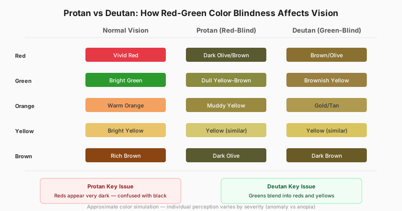
Protanopia vs. deuteranopia: both lose red-green discrimination, but a deuteranope's reds keep normal brightness — only the hue blurs.
What Deuteranopia Actually Is
Human color vision relies on three cone types. The M-cone peaks in sensitivity around 534 nm (medium-wavelength, green light). When the OPN1MW gene that encodes the M-cone photopigment is missing or severely altered, that cone is not produced. The remaining L-cone and S-cone still work, but the brain loses the ability to compare L and M signals — which is exactly what tells red from green. The result: a ripe red strawberry on a green bush looks like one uniform color.
Deuteranopia vs. Deuteranomaly — Where the Line Is
These two are often used interchangeably, but they are the two ends of one axis. Deuteranomaly still has M-cones — they are just shifted toward red — so color discrimination is only reduced. It is the mildest and by far the most common form of color blindness, affecting about 1 in 20 men, and many never find out.
Deuteranopia has no M-cone at all, so an entire axis of red-green discrimination is gone. Deuteranomaly is "colors shifted a little"; deuteranopia is "a whole dimension missing." Both get lumped under "deutan deficiency," but the severity gap is large: deuteranomalous people often pass most everyday color tasks, while deuteranopes reliably see red and green as the same color.
How It Affects Everyday Vision
A deuteranope does not see the world in gray — blues and yellows stay vivid. What collapses is the red-green axis. Because reds keep normal brightness, it is much quieter than protanopia: there is no dramatic loss of red signals, so many deuteranopes only realize something is off when someone points it out or they fail a screening. What genuinely trips them up are tasks that depend on telling red from green — red-green charts, judging ripeness, status indicator lights.
Causes and Genetics
Deuteranopia is almost always inherited. The OPN1MW gene sits on the X chromosome (locus Xq28) and is passed down in an X-linked recessive pattern. Males have only one X chromosome, so a single defective copy causes the condition; females need both copies defective — which is why deuteranopia is overwhelmingly seen in men. If the mother is a carrier, each son has a 50% chance of being deuteranopic. For a deeper look at inheritance, see our genetics of color blindness guide.
How Deuteranopia Is Diagnosed
- Ishihara test — fast screen for red-green deficiency; flags deuteranopia but cannot separate deutan from protan on its own.
- Red-green color blind test — targeted screen of the red-green axis, good for a quick first self-check.
- Farnsworth D-15 test — an arrangement test that reveals the deutan confusion axis and separates deutan from protan.
- FM100 Hue test — the most detailed arrangement test; identifies type and severity.
- Anomaloscope — the clinical gold standard, done at an eye clinic.
When to See a Doctor
Inherited deuteranopia is stable, lifelong, and needs no treatment in itself. But if your color vision changes suddenly, affects only one eye, or comes with reduced visual acuity, see an eye-care professional promptly — that pattern can point to a retinal or optic-nerve disease rather than inherited color blindness.
Daily Life Impact (5 Concrete Scenarios)
- Red-green charts — data visualizations and transit maps are the classic trap.
- Judging produce — red fruit among green leaves, or green vs. red peppers.
- Status indicators — red/green device LEDs (ready vs. error) are hard to tell apart.
- Matching colors — confusing green with brown, or pink with gray, when picking clothes or paint.
- Nature — the color depth of autumn leaves, lawns, and flowerbeds flattens out.
Tips and Adaptation
- Use labels, patterns, and position instead of red-green color coding alone.
- A phone color-picker app can name an object's true color on the spot.
- OS-level colorblind filters and high-contrast modes improve on-screen reading.
- Choose colorblind-safe palettes (Wong, Viridis) over red-green contrasts.
- Corrective glasses (e.g. EnChroma) help some people but vary — see how color blind glasses work.
Frequently Asked Questions
Sources
- National Eye Institute (NEI) — Overview of color blindness types and prevalence
- American Academy of Ophthalmology (AAO) — Clinical information on red-green color vision deficiency
- Colour Blind Awareness — Deuteranopia confused-color list and inheritance
- National Health Service (NHS) — Causes and management of colour vision deficiency
Take the Red-Green Test
Screen your red-green vision in 2 minutes
View Simulation
See the world through deuteranopia
Related Types
- Protanopia (Red-Blind) — red-green blindness with missing L-cones; reds are additionally darkened
- Tritanopia (Blue-Blind) — rare blue-yellow deficiency, often acquired
- All 8 types of color blindness — overview